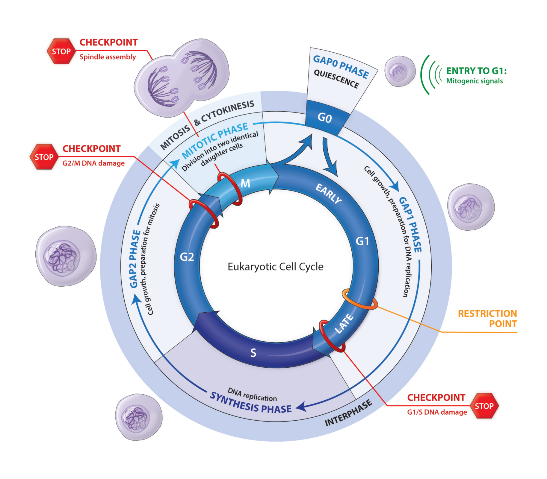
The diagram shows the stages of the eukaryotic cell cycle. – The eukaryotic cell cycle, a fascinating dance of cellular life, unfolds in a series of intricate stages, each playing a crucial role in the growth, division, and destiny of cells. The diagram accompanying this article serves as a visual guide, charting the key events that orchestrate this remarkable process.
From the bustling activities of interphase to the dramatic choreography of mitosis and cytokinesis, we will delve into the intricacies of the cell cycle, exploring the molecular machinery and checkpoints that ensure its precise execution. Along the way, we will uncover the practical applications of cell cycle analysis in medicine and research, shedding light on its significance in our understanding of life’s fundamental processes.
Overview of the Eukaryotic Cell Cycle

The eukaryotic cell cycle is a continuous process of growth and division that occurs in eukaryotic cells. It consists of four distinct stages: interphase, prophase, metaphase, anaphase, and telophase. During interphase, the cell grows and prepares for division. During prophase, the chromosomes become visible and the nuclear envelope begins to break down.
If you’re worried about your tires being stolen, there are a few things you can do to prevent it. Here are some tips to keep your tires safe.
During metaphase, the chromosomes line up in the center of the cell. During anaphase, the chromosomes are separated and pulled to opposite ends of the cell. During telophase, two new nuclear envelopes form around the chromosomes and the cell membrane pinches in the middle, dividing the cell into two daughter cells.
Interphase
Interphase is the longest stage of the cell cycle and is divided into three sub-phases: G1, S, and G2. During G1, the cell grows and prepares for DNA replication. During S, the cell’s DNA is replicated. During G2, the cell checks for DNA damage and prepares for mitosis.
Prophase, The diagram shows the stages of the eukaryotic cell cycle.
Prophase is the first stage of mitosis. During prophase, the chromosomes become visible and the nuclear envelope begins to break down. The centrosomes, which are responsible for organizing the spindle fibers, begin to move to opposite ends of the cell.
If you’re wondering how long your dishwasher cycle is, it can vary depending on the type of cycle you choose. This article provides an overview of the different dishwasher cycles and their approximate run times.
Metaphase
Metaphase is the second stage of mitosis. During metaphase, the chromosomes line up in the center of the cell. The spindle fibers attach to the chromosomes and begin to pull them apart.
Anaphase
Anaphase is the third stage of mitosis. During anaphase, the chromosomes are separated and pulled to opposite ends of the cell. The spindle fibers shorten, pulling the chromosomes apart.
Telophase
Telophase is the fourth and final stage of mitosis. During telophase, two new nuclear envelopes form around the chromosomes and the cell membrane pinches in the middle, dividing the cell into two daughter cells.
Interphase
Interphase is the longest phase of the cell cycle, occupying about 90% of its duration. During interphase, the cell grows, replicates its DNA, and prepares for cell division. Interphase consists of three distinct phases: G1, S, and G2.
G1 Phase
The G1 phase, also known as the first gap phase, is the first and longest phase of interphase. During the G1 phase, the cell grows and increases in size. It also synthesizes new proteins and organelles. The G1 phase is regulated by a checkpoint called the G1 checkpoint.
The G1 checkpoint ensures that the cell is ready to enter the S phase.
S Phase
The S phase, also known as the synthesis phase, is the second phase of interphase. During the S phase, the cell replicates its DNA. The DNA is replicated in a semi-conservative manner, meaning that each new DNA molecule consists of one original strand and one newly synthesized strand.
The S phase is regulated by a checkpoint called the S checkpoint. The S checkpoint ensures that the DNA is replicated accurately.
G2 Phase
The G2 phase, also known as the second gap phase, is the third and final phase of interphase. During the G2 phase, the cell prepares for cell division. It synthesizes new proteins and organelles, and it checks for DNA damage.
The G2 phase is regulated by a checkpoint called the G2 checkpoint. The G2 checkpoint ensures that the cell is ready to enter mitosis.
Mitosis
Mitosis is the process of cell division that results in two genetically identical daughter cells. It is divided into four stages: prophase, metaphase, anaphase, and telophase.
Prophase, The diagram shows the stages of the eukaryotic cell cycle.
Prophase is the first and longest stage of mitosis. During prophase, the chromosomes become visible and the nuclear envelope begins to break down. The spindle fibers, which are responsible for separating the chromosomes, begin to form.
Metaphase
Metaphase is the second stage of mitosis. During metaphase, the chromosomes line up in the center of the cell. The spindle fibers attach to the chromosomes and begin to pull them apart.
Anaphase
Anaphase is the third stage of mitosis. During anaphase, the chromosomes continue to be pulled apart by the spindle fibers. The chromosomes reach opposite ends of the cell.
Telophase
Telophase is the fourth and final stage of mitosis. During telophase, the spindle fibers disappear and the nuclear envelope reforms around each of the two daughter cells. The chromosomes become less visible and the cell begins to divide into two individual cells.
Cytokinesis
Cytokinesis is the final stage of the cell cycle, during which the cytoplasm divides, resulting in the formation of two daughter cells. It follows mitosis, where the chromosomes have been separated and packaged into two new nuclei.
Cytokinesis occurs differently in animal and plant cells due to the presence or absence of a cell wall. In animal cells, a cleavage furrow forms, pinching the cell membrane inward until the cell is completely divided. In plant cells, a cell plate forms in the center of the cell, eventually dividing the cell into two compartments.
Division of the Cell Membrane and Organelles
During cytokinesis, the cell membrane and organelles are also divided between the two daughter cells. In animal cells, the cell membrane is pinched inward by microfilaments made of the protein actin. In plant cells, the cell plate grows from the center of the cell outward, eventually fusing with the existing cell membrane to divide the cell.
Organelles are also divided during cytokinesis. Some organelles, such as mitochondria and ribosomes, are simply distributed randomly between the two daughter cells. Other organelles, such as the Golgi apparatus and endoplasmic reticulum, are divided by pinching or fragmentation.
Regulation of Cytokinesis
Cytokinesis is regulated by a complex network of proteins and signaling pathways. These proteins and pathways ensure that cytokinesis occurs at the right time and in the right place.
- Cyclin-dependent kinases (CDKs)are a family of proteins that regulate the cell cycle. CDKs are activated by cyclins, which are proteins that are produced and degraded at specific times during the cell cycle. CDKs phosphorylate other proteins, which triggers the events of cytokinesis.
- Rho GTPasesare a family of proteins that regulate the actin cytoskeleton. Rho GTPases are activated by guanine nucleotide exchange factors (GEFs) and inactivated by GTPase-activating proteins (GAPs). Rho GTPases control the formation of the cleavage furrow in animal cells.
- Aurora kinasesare a family of proteins that regulate chromosome segregation and cytokinesis. Aurora kinases phosphorylate other proteins, which triggers the events of cytokinesis.
Regulation of the Cell Cycle

The cell cycle is a highly regulated process to ensure the accurate duplication and distribution of genetic material. This regulation involves various checkpoints and molecular mechanisms.
Cyclins and Cyclin-Dependent Kinases (CDKs)
Cyclins are proteins that fluctuate in concentration throughout the cell cycle. They bind to and activate cyclin-dependent kinases (CDKs), which are enzymes that phosphorylate specific target proteins. The cyclin-CDK complexes control the progression of the cell cycle by regulating the activity of these target proteins.
Checkpoints
Checkpoints are control points in the cell cycle where the cell assesses whether it is ready to proceed to the next stage. There are three main checkpoints:* G1 checkpoint:Ensures the cell is ready to enter the S phase, where DNA replication occurs.
G2/M checkpoint
Ensures the cell is ready to enter mitosis, where the chromosomes are separated and distributed into two daughter cells.
M checkpoint (spindle assembly checkpoint)
Ensures all chromosomes are properly attached to the spindle fibers before anaphase begins.
Disruption of Cell Cycle Regulation
Disruption of cell cycle regulation can lead to various problems, including:* Cancer:Uncontrolled cell division can lead to the formation of tumors.
Developmental abnormalities
Errors in cell cycle regulation can cause birth defects.
Neurodegenerative diseases
Cell cycle dysregulation has been implicated in Alzheimer’s disease and Parkinson’s disease.
Applications of the Cell Cycle
Cell cycle analysis has a wide range of applications in medicine and research. It plays a crucial role in diagnosing and treating diseases like cancer and in developing new drugs.
One of the most important applications of cell cycle analysis is in the diagnosis and treatment of cancer. Cancer cells often have abnormal cell cycles, characterized by uncontrolled cell division. By analyzing the cell cycle of cancer cells, doctors can determine the stage and aggressiveness of the cancer, which helps guide treatment decisions.
Use of Cell Cycle Inhibitors in Drug Development
Cell cycle inhibitors are drugs that block specific cell cycle checkpoints, preventing cells from dividing. These drugs are being developed to treat cancer by targeting the uncontrolled cell division that characterizes cancer cells. By inhibiting cell cycle progression, these drugs can prevent cancer cells from proliferating and spreading.
Conclusion: The Diagram Shows The Stages Of The Eukaryotic Cell Cycle.
The eukaryotic cell cycle stands as a testament to the intricate symphony of life, a carefully orchestrated process that ensures the continuity and integrity of all living organisms. By unraveling its complexities, we gain a deeper appreciation for the remarkable dance of cellular renewal and division, a dance that lies at the heart of life’s grand tapestry.
Questions and Answers
What is the significance of the cell cycle?
The cell cycle is essential for the growth, development, and reproduction of all living organisms. It ensures the orderly duplication and distribution of genetic material, allowing cells to divide and create new cells.
What are the key stages of the eukaryotic cell cycle?
The eukaryotic cell cycle consists of two main phases: interphase and the mitotic phase. Interphase includes the G1, S, and G2 phases, during which the cell grows, synthesizes DNA, and prepares for division. The mitotic phase includes mitosis, where the chromosomes are segregated and divided, and cytokinesis, where the cytoplasm is divided.
How is the cell cycle regulated?
The cell cycle is tightly regulated by a complex network of proteins, including cyclins and cyclin-dependent kinases (CDKs). These proteins ensure that the cell cycle progresses in an orderly manner and that checkpoints are in place to prevent errors.
